
We employ techniques from astrophysics and astroparticle physics to enhance, develop and apply X-ray imaging methods and to investigate dosimetry applications.
Interferometric X-ray imaging
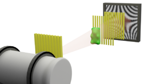
Conventional X-ray imaging operates on the principle that different materials absorb X-rays to varying degrees. Interferometric X-ray imaging is an extended imaging technique which leads besides the common attenuation image to two supplementary types of images: The differential phase-contrast and the dark-field image. By inserting microstructured gratings in a conventional X-ray imaging setup, the phaseshift imprinted on an X-ray wavefront by a sample, is analyzable. The differential phase-contrast image enhances differences between light elements, while the dark-field image is particularly sensitive to small angle scattering at tiny porous or fibrous structures, even on subpixel scale.
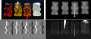
In Figure 2 four gummy bears are shown prepared with different objects (wooden toothpick, metal needle, micrometre-sized powder) to visualize the advantages of each obtained image. The metal needle is best visible in the attenuation image. Wood and powder are clearly visible in the dark-field image. The differential phase image is sensitive to edges and shows finer details of the gummy bear shape and structure.
We contribute to the development of the imaging method to further improve the feasibility of interferometric X-ray imaging. Additionally, we evaluate the potential of X-ray phase-contrast and dark-field imaging for different fields of application like medical imaging, non-destructive testing and laboratory astrophysics.